Two-organ on-a-chip device reproduces acetaminophen metabolism

gi1_00Mahler, G.J., Esch M.B., Shuler, M.L. “Characterization of a Gastrointestinal Tract Microscale Cell Culture Analog Used to Predict Drug Toxicity ”, Biotechnology & Bioengineering, 2009, 104(1), 193-205.

The oral uptake of acetaminophen was simulated with a two-organ microfluidic cell culture platform. The platform contained tissues that mimicked the GI tract epithelium and the liver. Concentrations of metabolites of acetaminophen in the cell culture medium were monitored over 24 hours, revealing that the system was capable of reproducing the uptake and first pass metabolism of acetaminophen.

LOC 2014: A two-organ system is more sensitive to damage by nanoparticles than single tissues in isolation

LOC ribbon-01Mandy B. Esch, Gretchen J. Mahler, Tracy Stokol, Michael L. Shuler, Body-on-a-Chip simulation with gastrointestinal and liver tissue suggests that ingested nanoparticles have the potential to cause liver injuryLab on a Chip, 2014, 14, 3081-3092.

In this paper we present a microfluidic two-organ system with GI tract tissue and liver tissue that interact with each other through soluble metabolite exchange. The results obtained with this system suggest that two-organ systems can detect toxicity at lower concentrations of 50 nm carboxylated nanoparticles than either of the two in vitro tissues alone. In addition, the GI tract epithelium filters out nanoparticle aggregates, preventing them from entering the systemic circulation and allowing passage to single nanoparticles only.

BMES 2014 reviewer’s choice award: Primary liver cells elevate metabolism in pumpless microfluidic device

blue ribbon-01

Mandy B. Esch, Jean Prot, Ying Wang, Paula Miller, Micheal L. Shuler

Introduction: Toxicity to the liver is one of the most common reasons for drug attrition during clinical trials. Liver cell culture devices that increase the sensitivity of liver cells to drug actions could help in identifying drugs that are toxic more easily. To decrease the risk posed to humans and animals, tests with in vitro tissues precede trials with animals, and tests with animals precede clinical trials with humans. Despite extensive testing, one cannot always predict drug toxicity accurately because animals and in vitro tissues do not recapitulate human tissues and metabolism as accurately as we need it. In this presentation we will discuss a liver cell culture device that could help improve early stage drug testing and also contribute to clarifying the mechanisms that enable hepatocytes to increase their metabolism in response to fluidic cell culture conditions.

Results: To create the fluidic flow in our devices in an inexpensive manner, we used a rocking platform that tilts the device at angles of ±12°, resulting in a periodically changing hydrostatic pressure drop and bidirectional fluidic flow (average flow rate of 650 µL min-1, and a maximum shear stress of 0.64 dyn min-2). We tested the performance of this cell culture device by co-culturing human primary non-parenchymal cells (fibroblasts, stellate, and Kupffer cells) with human primary hepatocytes for 14 days, finding that hepatocytes produced albumin and urea at elevated levels compared to static cultures. This result confirms our hypothesis that, similar to unidirectional flow, periodically changing bidirectional flow enhances the metabolic activity of hepatocytes. Hepatocytes also responded with P450 (CYP1A1 and CYP3A4) enzyme activity when challenged with P450 inducers throughout the 21 days of device operation. Non-parenchymal cells were similarly responsive, producing interleukin 8 (IL-8) when challenged with 10 µM bacterial lipoprotein (LPS). Flow rates were passively controlled via the dimensions of the microfluidic channels. Our results indicate that device operation with bi-directoinal gravity-driven medium flow supports the long-term culture of primary human liver cells with the benefits of enhanced metabolic activity. Our mode of device operation allows us to evaluate drugs under fluidic cell culture conditions and at low device manufacturing and operation costs.

How body-on-a-chip devices can help foster drug development: an outline of strategies

Microphysiologic Cell Culture Systems are microfluidic platforms on which multiple in vitro tissues can communicate with each other via soluble metabolites that re-circulate through a medium stream. The systems are also referred to as body-on-a-chip systems or micro cell culture analogs (µCCAs).

My research combines tissue engineering and microfabrication to construct microphysiological systems and to examine how cooperative interactions of organs result in the overall functioning of the human body. The collective effects of inter-organ metabolic exchanges are not only directly relevant to evaluating new drugs, they also raise a number of fascinating Systems Biology questions concerning the mechanisms of communication between organs that lead to the overall response of the human body to chemical and biological challenges.

Here is a paper that reviews some of the literature regarding micro physiological systems and outlines their possibilities for research:

Mandy B. Esch, Alec Smith, Jean-Mathew Prot, Charlotta Oleaga Sancho, James Hickman, Michael L. Shuler, How Multi-Organ Microdevices Can Help Foster Drug Development. Advanced Drug Delivery Reviews, 201469/70, 158-169.

Here are two papers that present a micro physiological system with GI tract  and liver tissues:

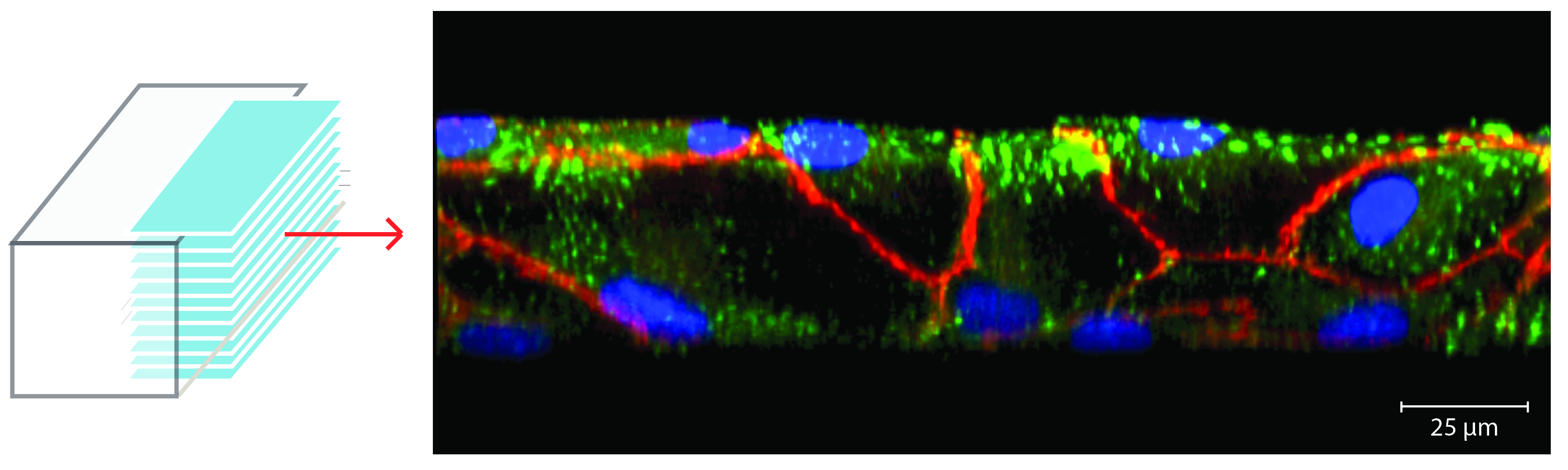
Mandy B. Esch, Gretchen J. Mahler, Michael L. Shuler, Body-on-a-Chip simulation with gastrointestinal and liver tissue suggests that ingested nanoparticles have the potential to cause liver injury. Lab on a Chip, 2014, 14, 3081-3092. doi: 10.1039/c41c00371c

Mahler, G.J., Esch M.B., Shuler, M.L. “Characterization of a Gastrointestinal Tract Microscale Cell Culture Analog Used to Predict Drug Toxicity ”, Biotechnol & Bioeng, 2009, 104(1), 193-205.

3D tissue scaffold for on-chip gut: microfabrication and integration into fluidic cell culture platform

Image7-279jrsn.jpg

Esch, M.B., Sung, J.H., Yang, J. Yu, J., March, J.C., Shuler, M.L. “On Chip Porous Polymer Membranes for Integration of Gastrointestinal Tract Epithelium with Microfluidic ‘Body-on-a-Chip’ Devices”, Biomedical Microdevices, 2012, 14 (5), 895-906.

We have developed a method to fabricate porous polymer membranes that are shaped in 3D to form 50 x 50 μm tall villi. This membrane was used as a scaffold to culture gut cells (Caco-2 and primary gut cells) on silicon within microfluidic systems. The 3D cell cultures can be used to simulate the oral uptake and absorption of nutrients, drugs, and drug carriers.  When incorporated into a microfluidic system that facilitates the recirculation of medium the tissue can also be combined with other on-chip tissues such as liver tissue to simulate the first pass metabolism. First pass metabolism refers to the absorption of ingested substances through intestinal tissue and their immediate transfer to the liver where enzymes can metabolize them before they enter the systemic circulation. The process considerably decreases the bioavailability of ingested substances and is of interest to drug developers, toxicologists, and nutrition scientists.

Nature Nanotech: nanoparticles influence iron uptake and change the sizes of macrovilli in the gut

Gretchen J. Mahler, Mandy B. Esch, Elad Tako, Shivaun D. Archer, Raymond P. Glahn, Michael L. Shuler, Oral Exposure to Nanoparticles Affects Essential Nutrient Absorption.  Nature Nanotechnology, 2012, 7, 264–271  (this publication was covered by the MRS bulletin and Science Daily.)

This paper describes the effects of nanoparticle injestion on the GI tract epithelium. Using in vitro analysis and in vivo models (chicken), we found that nanoparticle injestion affects nutrient (iron) uptake negatively, and that the body compensates in the long term by increasing the surface of the its GI tract epithelium. This study looks at non-lethal, long-term effects of nanoparticles, finding that real physiologic consequences arise from nanoparticle injestion.

Tissue Engineering A: 50 micrometer in vitro microvasculature (endothelial lining) establishes adherents junctions even on the sidewalls of channels

microvasculature results 2-01Esch, M.B., Post, D., Shuler, M.L., Stokol, T. “Characterization of Small Diameter In Vitro Endothelial Linings of the Microvasculature,” Tissue Engineering A, 2011, 17, 2965-2971

In vitro microvascular endothelial linings cultured in 50 µm wide, 50 µm high microfluidic vessels establish tight junctions that allow us to use them to investigate biophysical and molecular mechanisms that play a role in circulatory disease phenomena. In vivo, endothelial cells grow on the inner surface of blood vessels and are confined by its geometry. In the smallest vessels of the microvasculature, this confinement leads to a significant bend within each cell. To imitate these geometric constraints within an in vitro model of the endothelial lining, we have fabricated small microfluidic channels (50 µm wide, 50 µm high) and cultured human umbilical vein endothelial cells (HUVECs) within them. We have characterized the developed model and our results show that the cells are capable of establishing adherents junctions (shown in the picture in red) even at the sidewalls of the channels.

Small vessels fully lined with endothelial cells can be used to study circulatory diseases

Mandy B. Esch, David J. Post, Michael L. Shuler, Tracy Stokol, Characterization of Small Diameter In Vitro Endothelial Linings of the Microvasculature. Tissue Engineering A, 2011, 17, 2965-2971

In this paper we present ultra-small microfluidic vessels (50 x 50 µm in cross section) that are fully lined with endothelial cells. We used confocal imaging to create 3D views, confirming that not only the top and bottom walls of the channels are fully lined with cells, but also the sidewalls. Fully established barrier functions are necessary in order to use the vessels for drug uptake studies. In addition, control over the barrier function is necessary for studies that aim to find the cause of circulating tumor cells attachment to the endothelium.

Analytical Chemistry: Microfluidic sensor for sensing heat-shock mRNA of Cryptosporidium parvum

Esch, M.B.; Locascio, L.; Tarlov, M.J.; Durst, R.A. “Detection of Viable Cryptosporidium parvum in a Microfluidic Chip”, Analytical Chemistry, 2001, 73 (13), 2952-2958.Crypto

In this project we developed a microfluidic RNA sensor that improved sensitivity compared to previously developed injection flow analysis-based sensors 5-fold. The sensor captured RNA from Cryptosporidium parvum within a microfluidic channel. Subsequent flow of oligonucleotide-tagged fluorescent green dye-filled liposomes made the RNA visible within the microfluidic channel. This work was presented at Pittcon in 2001.


Pittcon: Paper-based microfluidics for the detection of C. parvum mRNA in 30 min

Esch, M.B.; Bäumner, A.; Durst, R.A. “Rapid Visual Detection of Viable Cryptosporidium parvum on Test Strips using Oligonucleotide-tagged Liposomes” Analytical Chemistry, 2001, 73(13), 3162-3167

Paper-based microfluidics are the basis for pregnancy test strips that detect hormones via antibodies that are immobilized on a paper test strip. We employed the same principle to detect the RNA of a pathogen (Cryptosporidium parvum).  We immobilized synthetic DNA that is complementary to the RNA and placed it on the test strip. While the sample moves up the strip via capillary forces, the RNA in the sample comes into very close contact with the immobilized DNA. In our assay, the RNA competes with a second dye-conjugated synthetic DNA with the same sequence. This dye-conjugated DNA was initially added to the sample. In this competitive assay format, no color on the strip means the pathogen RNA was present.

Biopolymers: Relaxation of water confirms experimentally that water is bound to hydrogels via non-covalent forces

rotationschematic2-01

Esch, M.B.; Sukhorukov, V.L.; Kürschner, M.; Zimmermann, U. “Dielectric Properties of Alginate Beads and Bound Water Relaxation Studied by Electrorotation” Biopolymers, 1999, 50, 227-237

The association of water with polysaccharides is the basis for the survival of cells within hydrogels. Hydrogel-encapsulated cells are important because they could replace native cells that have experienced a loss of function, for example insensitivity to insulin-needs of the body. Here we proved experimentally that water is bound non-covalently to hydrogels. To prove this, we used four microelectrodes to create a rotating electric field in which we placed 400 micrometer large alginate beads. We measured how fast the beads rotated in fields of varying frequency and in medium of varying conductivity. A broad internal dispersion of the hydrogel centered between 20 and 40 MHz. We attribute this dispersion to the relaxation of water bound to the polysaccharide matrix of the beads.