 Esch, M.B., Post, D., Shuler, M.L., Stokol, T. “Characterization of Small Diameter In Vitro Endothelial Linings of the Microvasculature,” Tissue Engineering A, 2011, 17, 2965-2971
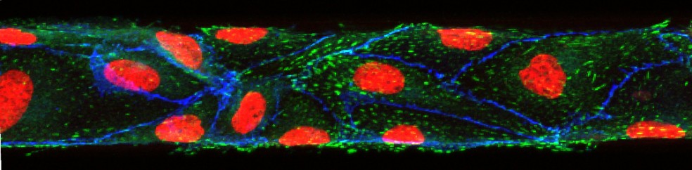
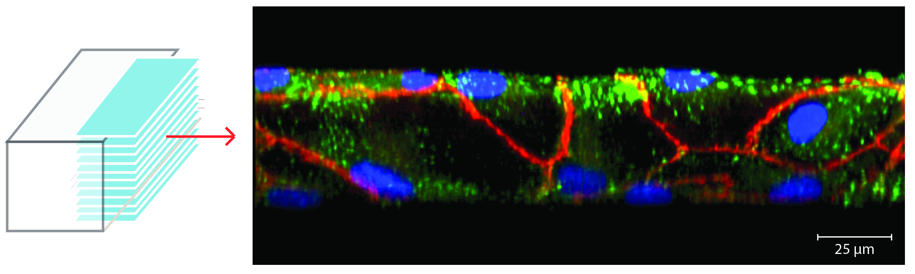
In vitro microvascular endothelial linings cultured in 50 µm wide, 50 µm high microfluidic vessels establish tight junctions that allow us to use them to investigate biophysical and molecular mechanisms that play a role in circulatory disease phenomena. In vivo, endothelial cells grow on the inner surface of blood vessels and are confined by its geometry. In the smallest vessels of the microvasculature, this confinement leads to a significant bend within each cell. To imitate these geometric constraints within an in vitro model of the endothelial lining, we have fabricated small microfluidic channels (50 µm wide, 50 µm high) and cultured human umbilical vein endothelial cells (HUVECs) within them. We have characterized the developed model and our results show that the cells are capable of establishing adherents junctions (shown in the picture in red) even at the sidewalls of the channels.
