Two-organ on-a-chip device reproduces acetaminophen metabolism

gi1_00Mahler, G.J., Esch M.B., Shuler, M.L. “Characterization of a Gastrointestinal Tract Microscale Cell Culture Analog Used to Predict Drug Toxicity ”, Biotechnology & Bioengineering, 2009, 104(1), 193-205.

The oral uptake of acetaminophen was simulated with a two-organ microfluidic cell culture platform. The platform contained tissues that mimicked the GI tract epithelium and the liver. Concentrations of metabolites of acetaminophen in the cell culture medium were monitored over 24 hours, revealing that the system was capable of reproducing the uptake and first pass metabolism of acetaminophen.

LOC 2014: A two-organ system is more sensitive to damage by nanoparticles than single tissues in isolation

LOC ribbon-01Mandy B. Esch, Gretchen J. Mahler, Tracy Stokol, Michael L. Shuler, Body-on-a-Chip simulation with gastrointestinal and liver tissue suggests that ingested nanoparticles have the potential to cause liver injuryLab on a Chip, 2014, 14, 3081-3092.

In this paper we present a microfluidic two-organ system with GI tract tissue and liver tissue that interact with each other through soluble metabolite exchange. The results obtained with this system suggest that two-organ systems can detect toxicity at lower concentrations of 50 nm carboxylated nanoparticles than either of the two in vitro tissues alone. In addition, the GI tract epithelium filters out nanoparticle aggregates, preventing them from entering the systemic circulation and allowing passage to single nanoparticles only.

BMES 2014 reviewer’s choice award: Primary liver cells elevate metabolism in pumpless microfluidic device

blue ribbon-01

Mandy B. Esch, Jean Prot, Ying Wang, Paula Miller, Micheal L. Shuler

Introduction: Toxicity to the liver is one of the most common reasons for drug attrition during clinical trials. Liver cell culture devices that increase the sensitivity of liver cells to drug actions could help in identifying drugs that are toxic more easily. To decrease the risk posed to humans and animals, tests with in vitro tissues precede trials with animals, and tests with animals precede clinical trials with humans. Despite extensive testing, one cannot always predict drug toxicity accurately because animals and in vitro tissues do not recapitulate human tissues and metabolism as accurately as we need it. In this presentation we will discuss a liver cell culture device that could help improve early stage drug testing and also contribute to clarifying the mechanisms that enable hepatocytes to increase their metabolism in response to fluidic cell culture conditions.

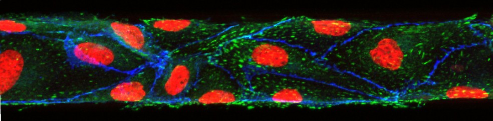
Results: To create the fluidic flow in our devices in an inexpensive manner, we used a rocking platform that tilts the device at angles of ±12°, resulting in a periodically changing hydrostatic pressure drop and bidirectional fluidic flow (average flow rate of 650 µL min-1, and a maximum shear stress of 0.64 dyn min-2). We tested the performance of this cell culture device by co-culturing human primary non-parenchymal cells (fibroblasts, stellate, and Kupffer cells) with human primary hepatocytes for 14 days, finding that hepatocytes produced albumin and urea at elevated levels compared to static cultures. This result confirms our hypothesis that, similar to unidirectional flow, periodically changing bidirectional flow enhances the metabolic activity of hepatocytes. Hepatocytes also responded with P450 (CYP1A1 and CYP3A4) enzyme activity when challenged with P450 inducers throughout the 21 days of device operation. Non-parenchymal cells were similarly responsive, producing interleukin 8 (IL-8) when challenged with 10 µM bacterial lipoprotein (LPS). Flow rates were passively controlled via the dimensions of the microfluidic channels. Our results indicate that device operation with bi-directoinal gravity-driven medium flow supports the long-term culture of primary human liver cells with the benefits of enhanced metabolic activity. Our mode of device operation allows us to evaluate drugs under fluidic cell culture conditions and at low device manufacturing and operation costs.

Nature Nanotech: nanoparticles influence iron uptake and change the sizes of macrovilli in the gut

Gretchen J. Mahler, Mandy B. Esch, Elad Tako, Shivaun D. Archer, Raymond P. Glahn, Michael L. Shuler, Oral Exposure to Nanoparticles Affects Essential Nutrient Absorption.  Nature Nanotechnology, 2012, 7, 264–271  (this publication was covered by the MRS bulletin and Science Daily.)

This paper describes the effects of nanoparticle injestion on the GI tract epithelium. Using in vitro analysis and in vivo models (chicken), we found that nanoparticle injestion affects nutrient (iron) uptake negatively, and that the body compensates in the long term by increasing the surface of the its GI tract epithelium. This study looks at non-lethal, long-term effects of nanoparticles, finding that real physiologic consequences arise from nanoparticle injestion.

Small vessels fully lined with endothelial cells can be used to study circulatory diseases

Mandy B. Esch, David J. Post, Michael L. Shuler, Tracy Stokol, Characterization of Small Diameter In Vitro Endothelial Linings of the Microvasculature. Tissue Engineering A, 2011, 17, 2965-2971

In this paper we present ultra-small microfluidic vessels (50 x 50 µm in cross section) that are fully lined with endothelial cells. We used confocal imaging to create 3D views, confirming that not only the top and bottom walls of the channels are fully lined with cells, but also the sidewalls. Fully established barrier functions are necessary in order to use the vessels for drug uptake studies. In addition, control over the barrier function is necessary for studies that aim to find the cause of circulating tumor cells attachment to the endothelium.